HISTOLOGICAL EVALUATION OF HUMAN PULP TISSUE IN RESPONSE TO CARIES PROGRESSION
EVALUASI HISTOLOGIS RESPON JARINGAN PULPA GIGI MANUSIA TERHADAP TINGKAT KEPARAHAN KARIES
DOI:
https://doi.org/10.32734/dentika.v15i2.2004Keywords:
dental pulp, caries, histological evaluationAbstract
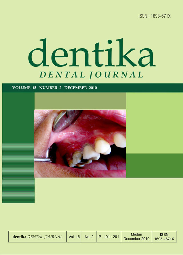
The dental pulp is protected by enamel, dentin, and cementum from the microbial rich oral environment. Carious lesion may provide pathways for microorganisms and their toxins to enter the pulp. This study aimed to evaluate the responses of human dental pulp to caries progression. Twenty volunteers who had been scheduled to undergo extraction for various therapeutic reasons, were enrolled in the study. Five intact third molars and 15 third molars with carious lesion involving enamel, dentin, and pulp were extracted. Before decalcifying with 10% EDTA solution (pH 7.4), the lesion conditions were confirmed with micro-computed tomography. The specimens were embedded in paraffin, sectioned serially and stained with Hematoxylin Eosin. The specimens were then evaluated under a light microscope. All normal intact teeth showed histological features of normal pulp tissue with no inflammation. All teeth with carious lesion confined to enamel revealed slight inflammation and disorganization of odontoblast layer under to the lesions. Most samples with caries confined to dentin showed slight inflammation, only 1 sample showed moderate inflammation. Odontoblast layer lost its continuity and could not be observed in the areas under to the lesions. Severe inflammation was observed in 3 out of 5 carious teeth with pulp exposure, while the remaining samples showed moderate inflammation. Numerous blood vessels were found in the tissue surrounding the intense cellular infiltration. In conclusion, caries may induce inflammation and disorganization of the pulp tissue. The severity of pulp inflammation and disorganization depends on the depth of the lesion.
Downloads